")
Strongyloides is a genus of nematodes from the Strongyloididae family. They affect the small intestine of a large number of vertebrate species, including domestic carnivores and primates. Different species have been described in non-human primates, namely Strongyloides fuelleborni, which is the most frequently encountered one, but also S. cebus, S. papillosus, S. simiae and S. stercoralis (Cogswell, 2007).
Epidemiology
Although Strongyloides parasites are more prevalent in subtropical and tropical regions, the parasite remains cosmopolitan and can also be found in temperate climates. They are found in all non-human primate groups. Different species of parasites have been reported to affect different species of animals:
- Strongyloides stercoralis in New World Monkeys, namely monk sakis (Pithecia monachus) (Rondón et al., 2021), Old-World Monkeys, namely proboscis monkeys (Nasalis larvatus) (Adrus et al., 2019), and Apes, namely gorillas (Gorilla), orangutans (Pongo spp.), gibbons (Hylobatidae) and chimpanzees (Pan spp.) (Cogswell, 2007; Medkour et al., 2020);
- Strongyloides fuelleborni in Prosimians, Old World Monkeys and Great Apes, namely in Bornean slow lorises (Nycticebus menagensis), rhesus macaques (Macaca mulatta), crab-eating macaques (Macaca fascicularis), pig-tailed macaques (Macaca nemestrina), baboons (Papio), vervet monkeys (Chlorocebus pygerythrus and Chlorocebus sabeus), Uzungwa red colobus (Procolobus gordonorum), guenons (Cercopithecidae) and chimpanzees (Pan spp.) (Cogswell, 2007; Anderson et al., 2012; Calle & Joslin, 2015 ; Frias et al., 2018; Tanchomnang et al., 2018 ; Barelli et al., 2019 ; Janwan et al., 2020 ; Cruz et al., 2021) ;
- Strongyloides papillosus in Old World Monkeys and Great Apes, namely in common patas monkeys (Erythrocebus patas), gorillas (Gorilla), orangutans (Pongo spp.), gibbons (Hylobatidae) and chimpanzees (Pan spp.) (Cogswell, 2007);
- Strongyloides simiae in Old World Monkeys, namely in common patas monkeys (Erythrocebus patas) and macaques (Macaca) (Cogswell, 2007);
- Strongyloides cebus in New World Moneys, namely in squirrel monkeys (Saimiri), marmosets (Callitrichidae), spider monkeys (Ateles spp.), capuchin monkeys (Sapajus and Cebus spp.) and wooly monkeys (Lagothrix spp.) (Cogswell, 2007; Mati, 2013; Parr, 2013).
Description
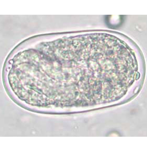
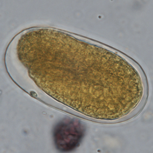
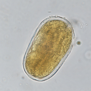
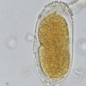
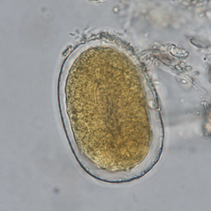
Strongyloides eggs are oval, symmetrical with parallel lateral sides. Their outer membrane is thin, and the eggs contain a vermiform embryo. They measure 40 to 70 µm long and 20 to 35 µm large. It is impossible to differentiate different species of Strongyloides using ovodiagnosis only. A coproculture is recommended to refine the diagnosis (Cogswell, 2007).
Differential diagnosis
Differential diagnosis includes parasitic and non-parasitic structures:
- Free-living nematodes: their eggs are typically larger than strongyle ones, ranging from 70 to 120 µm in length and 24 to 43 µm large. Moreover, some have typical characteristics like refringent globules in Heterodera radicicola. A coproculture or Baermann migration-based diagnosis are recommended in case of doubts (Petithory et al., 1995).
- Embryonated strongyle eggs: although they are approximately of the same size as Strongyloides eggs, they are asymmetrical. In case of doubts, it is recommended to carry out complementary exams like coproculture or Baermann migration method to refine the diagnosis (Garcia, 2021).
Clinical significance
Strongyloides infection causes dehydration, lethargy, weight-loss, anorexia, digestive clinical signs (mucoid-to-hemorrhagic diarrhea, vomiting, constipation), dermatological signs (dermatitis, pruritus), respiratory signs (coughing, dyspnea) and can even lead to death (Strait et al., 2012; Mati et al., 2013; Calle & Joslin, 2015; Murphy, 2015; Frias et al., 2018b; Cruz et al., 2021).
Prophylaxis and treatment
Strongyloidosis is highly contagious and some species like Strongyloides fuelleborni are zoonotic. Therefore, hygienic measures are key to manage the disease.
Various treatments have been described in non-human primates:
- Ivermectine: 200 to 300 µg/kg IM, SC, PO once (Calle & Joslin, 2015; Cruz et al., 2021);
- Thiabendazole: 50-100 mg/kg PO q24h during 1 to 5 days (Cogswell, 2007);
- Moxidectine: 0.5 mg/kg (Dufour et al., 2006);
- Mebendazole: 22 mg/kg PO or SC q24h during 2 to 3 days (Cogswell, 2007);
- Fenbendazole: 20 mg/kg PO q24h during 14 days (Calle & Joslin, 2015);
- Albendazole: 10 mg/kg PO q24h during 3 days (Cruz et al., 2021);
- Pyrantel: 11 mg/kg once (Strait et al., 2012);
- Levamisole: 10 mg/kg PO q24h during 2 to 3 days (Calle & Joslin, 2015).