Cyclospora is a genus of coccidial protozoa of the digestive tract of a large number of hosts (reptiles, insects, rodents, primates, other mammals). They are normally host-specific (Ortega & Eberhard, 2010). Various species have been described as pathogenic in non-human primates, namely Cyclospora cayetanensis, C. cercopitheci, C. colobi, C. papionis (Eberhard et al., 1999) and C. macacae (Li et al., 2015).
Epidemiology
Cyclospora spp. are cosmopolitan parasites found in Old and New World Monkeys, and Apes, namely in chimpanzees (Pan troglotydes), baboons (Papio spp.), vervet monkeys (Chlorocebus spp.), black-and-white colobus (Colobus spp.), macaques (Macaca spp.) and tamarins (Saguinus spp.) amongst other species (Eberhard et al., 1999; Strait et al., 2012; Li et al., 2015; Zapata Valencia et al., 2021). They may be transmissible to humans.
Description
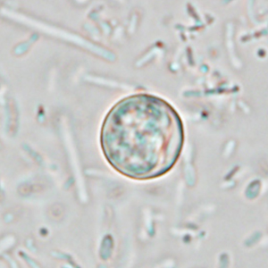
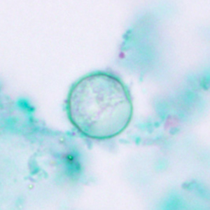
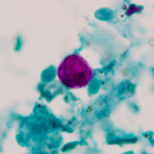
Oocysts visualized on fecal screening are round and measure between 8 and 10 µm in diameter. Both a sporulated and non-sporulated form exist. In sporulated cysts, 2 sporocyst each containing 2 sporozoites are present, but are difficult to visualize using optic microscopy (Colomina Rodriguez & Villar Serrano, 1997). Oocysts do not stain with Lugol but can be visualized using modified Ziehl-Neelsen staining. The final diagnosis can be made by the combination of three diagnostic modalities:
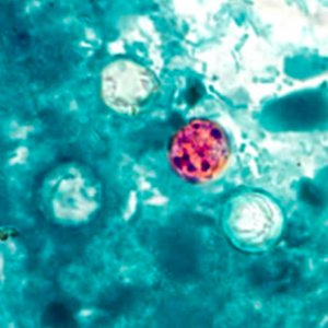
- Modified Ziehl Neelsen staining: oocysts are detected by transparency, or as red to pink cysts on a blue to green background (Strait et al., 2012);
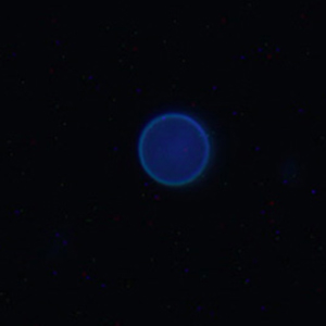
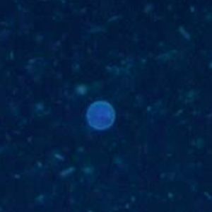
- Fluorescence microscopy: oocysts appear in blue when the wavelength is comprised between 330 and 365 nm (Li et al., 2015);
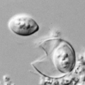
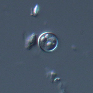
- Differential interference contrast microscopy: it allows the visualization of oocysts more distinctively (Verweij et al., 2003).
Differential diagnosis
The differential diagnosis includes other coccidial species, other small protozoan cysts and non-parasitic structures. However, small cysts of flagellates and amoebas (namely Retortamonas intestinalis, Enteromonas hominis, Endolimax nana, and Entamoeba hartmanni) often stain with Lugol stain and contain a nucleus. Like Cyclospora spp., other coccidial oocysts normally stain with modified Ziehl-Neelsen and not with Lugol. However, they often differ in size (4 to 6 µm for Crysptosporidium spp. and more than 10 µm for Eimeria and Isospora spp.) (Strait et al., 2012). Non-parasitic elements do not stain with neither modified Ziehl-Neelsen nor Lugol stain (Petithory et al., 1995).
Clinical significance
Cyclospora infection can cause digestive clinical signs, although these are poorly described in non-human primates. In humans, Cyclospora cayetanensis can be responsible for acute liquid diarrhea, abdominal pain, vomiting, anorexia, weight loss, myalgia, hyperthermia and lethargy (Global Health, Division of Parasitic Diseases and Malaria, 2019b).
Prophylaxis and treatment
As some Cyclospora species are zoonotic pathogens, hygienic measures need to be taken in case of diagnosis. In both humans and non-human primates, trimethoprim-sulfamethoxazole seem to be efficient in treating Cyclospora infections (Strait et al., 2012; Murphy, 2015).
")